Share this article and save a life!
AI just turned a routine MRI into a full body composition report. 🧠
No new scanner. No technologist manually measuring tissue slices. Five minutes.
AIRS Medical received FDA 510(k) clearance this week for SwiftSight Body Composition, an AI-powered platform that extracts quantitative body composition data directly from standard MR images. SimonMed Imaging is already lined up as the first customer to deploy it.
What it actually measures:
🔹 Visceral and subcutaneous fat
🔹 Abdominal and thigh muscle quantity, quality, and bilateral balance
🔹 Liver fat via proton density fat fraction (PDFF) with automated region-of-interest mapping
🔹 Percentile rankings benchmarked against age- and sex-matched reference populations
All of it delivered in a structured Body Composition Report, produced through automated AI segmentation, at scale, across multiple imaging locations.
The workflow angle is what grabs me.
BMI tells you almost nothing about metabolic risk. It cannot show where fat is distributed or how much functional muscle a patient actually has. According to the company, SwiftSight provides the granular picture that BMI has always been too blunt to deliver, and it does that inside an MRI scan a patient was already going to have.
That distinction matters in 2026. GLP-1 adoption is driving a surge of patients who need longitudinal body composition tracking. Weight on a scale tells you a drug is working. MRI tells you whether the patient is losing muscle or losing fat, which is a fundamentally different clinical question.
MRI does not use ionizing radiation. That means repeat exams over time are practical in a way CT-based body composition scans are not.
For imaging organizations, AIRS Medical is positioning this as a revenue expansion play, not just a clinical tool. High-volume MRI centers can add a differentiated quantitative service using infrastructure they already own, without a separate modality and without building a manual analysis workflow from scratch.
The roadmap signals ambition. According to AIRS Medical, diffusion tensor imaging is planned for November 2026, with quantitative joint applications to follow in 2027.
This is the pattern I keep watching: AI does not replace the MRI, it replaces the bottleneck between the image and the insight. The scanner was never the constraint. The time and expertise required to extract structured, reproducible, scalable information from those images, that was always the constraint.
Every modality in radiology is on a path toward this. The question worth sitting with is not whether AI can do the analysis. It is whether imaging organizations build that capability now or wait until every competitor in their market already has.
👉 Follow Jonathan Govette, CEO of Oatmeal Health, for daily healthcare insights on LinkedIn. Deeper dives in The Oatmeal Bite on Substack: https://news.oatmealhealth.com
Share this article and save a life!
Author:
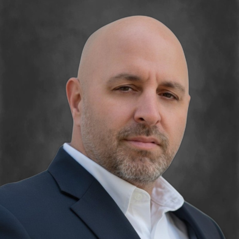
CEO/Co-Founder @ Oatmeal Health | AI Lung Cancer Screening | Almost Became a Doctor | Engineer | Follow to Share What I’ve Learned Along the Way
I help patients get the care they need earlier, preventing late-stage cancer.
That’s been the throughline across three companies and almost 20 years in healthcare. At ReferralMD, we fixed broken referral networks so patients didn’t fall through the cracks. At Oatmeal Health, it’s lung cancer: building the diagnostic and screening infrastructure so the 85% of cases caught too late get caught early instead.
Today as CEO of Oatmeal Health, I lead a team embedding AI into radiology workflows to turn routine lung CT scans into reimbursable cancer risk assessments. We partner with FQHCs to reach underserved communities, and with health systems and payers to make early detection economically sustainable. Think HeartFlow or Cleerly, but for lungs.
Between companies, I advised at Techstars and Plug and Play, mentoring founders building in digital health. That experience shaped how I think about what separates companies that ship from companies that stall: distribution, reimbursement, and clinical trust, not just technology.
I’m a CancerX alumnus, a 3x healthcare founder, and someone who believes the biggest problems in cancer aren’t scientific. They’re operational.
We’re hiring mission-driven builders at Oatmeal Health. If you want to work on something that matters, reach out.
When I’m not working, I’m traveling, mentoring, and keeping up with one very energetic husky. 🐾
Substack – The Oatmeal Bite:
Millions of patients get less care because of who they are, where they live, or how they look. I’m fighting to change that. CEO @OatmealHealth, a startup built for the underserved. The Oatmeal Bite: intel for clinicians, investors, and advocates.
Jonathan Govette
CEO of Oatmeal Health
Substack:
https://oatmealhealthjonathangovette.substack.com/